This post was originally published on this site
A U.S. Department of Health and Human Services grant will fund research at two universities into whether an ultrasound technology might be more effective than mammography at detecting breast cancer, particularly in younger people.
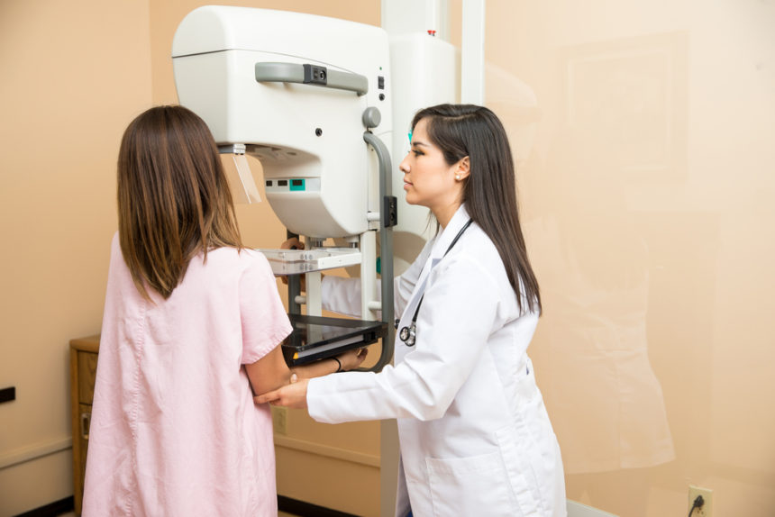
At present, mammography is most often used to screen for and diagnose breast cancers. However, mammograms are essentially X-rays, and aren’t especially good at detecting certain types of breast tumors, particularly in younger people who tend to have more dense breast tissue.
The new project will be headed by Mark Anastasio, PhD, a professor at the University of Illinois, and by Neb Duric, PhD, a professor at Wayne State University. That grant award is titled “Advanced image reconstruction for accurate and high-resolution breast ultrasound tomography.”
“The current methods of mammography … are based on X-rays,” Anastasio said in a press release. “We’re investigating this new technology that can be useful for breast cancer imaging that is based on the use of ultrasound instead of X-rays.”
In order to produce images, X-ray-based technologies use ionizing radiation — high-energy rays that can move through bodies to provide details about what’s inside. But these rays can also cause damage to cellular structures like DNA; that’s why X-ray technicians wear leaded aprons, which block X-rays.
The new technology will use ultrasound — sound waves — instead. These waves can also move through the body, but don’t carry the same safety risks: “Not only is it safer because it doesn’t involve ionizing radiation, it is more sensitive to certain tissue properties that will make it easier to detect subtle breast cancers,” said Anastasio.
Although some current imaging methods use ultrasounds, the images generated (via computational algorithms that translate the waves into an image) aren’t as clear as those made by X-ray-based techniques. This project aims to improve the clarity of these images so that they can be more effectively utilized in clinical settings.
“We quickly realized that through the use of advanced image reconstruction principles and high-performance computing, we can actually do a better job of modeling the physics and reconstruct images of much better quality,” Anastasio said. “They almost look like MRI images in terms of resolution.”
Anastasio also pointed out that this technology is particularly applicable to imaging the breast: “If you wanted to image a thicker part of the body, say the abdomen, it would be tougher because the ultrasound would be more attenuated [reduced] and more scattered.”
Over the four-year course of the grant, whose total was not specified, the researchers hope to better understand how ultrasounds interact with breast tissue, then use this information to inform the algorithms that develop images. Then, they will work with radiologists to figure out how best to translate these findings into clinical practice.
Once developed, the researchers hope that this technology could improve breast cancer detection, especially in younger people. Plus, it could be adapted to help determine an individual’s cancer risk.
“In the end, we hope to have a fully optimized and validated image construction algorithm where we can reconstruct 3D images of the breast that can be subsequently evaluated in large scale clinical trials and turned into a useful product,” Anastasio said.
The post HHS Grant to Advance Breast Cancer Screenings Based on Ultrasound appeared first on Breast Cancer News.
The post HHS Grant to Advance Breast Cancer Screenings Based on Ultrasound appeared first on BioNewsFeeds.


